Read on to learn about the insights that Surface Plasmon Resonance (SPR) can provide for immunotherapeutic research.
Introduction
According to John Hopkins Medicine, almost 40 percent of the population of the United States will be diagnosed with cancer during their lifetime. Despite this staggering statistic, the advancements being made in cancer research and treatments have dramatically changed the trajectory of patient lifecycles, with over 20 million people expected to live beyond a cancer diagnosis by 2026.1
Among the many innovations seen in the last decade, immunotherapy has emerged as one of the hottest areas of cancer research, with the promise of being a more effective and safer method of treating cancer, compared to more conventional therapies such as radiation and chemo.
Despite its success in treating certain cancers and its year-over-year development pipeline growth (which includes a 22 percent increase from 2019 to 2020), immunotherapy development has continued to be a challenging process due to the complex mechanism-of-action of the drugs and their immune-related targets.2
In this article, we discuss how bioassays such as surface plasmon resonance (SPR) can aid in the understanding of current immunotherapy targets and agents to develop more effective treatments against cancer.
Blog Overview
- What is immunotherapy
- Identifying and targeting biomarkers
- Understand the mechanism of immune escape
- Improving inefficiencies of monoclonal antibodies
- Advance your immunotherapy research with SPR
What is immunotherapy?
More than a decade ago, scientists made a critical discovery about tumour cells having the ability to evade or interfere with our immune systems, thus curbing our body’s natural response to abnormal cells that would otherwise target and destroy the tumour. Immunotherapy is a type of biological therapy that activates or harnesses our immune systems to better fight against invading tumour cells. It has been successfully applied in the prevention and even treatment of various forms of cancer.
Identifying and targeting biomarkers
In the field of oncology, immune-related biomarkers have helped in the discovery of specific or combinatorial immune therapies as potential therapeutic targets. Treatments can involve the use of antibodies, CAR-T cell therapy, cancer vaccines, adoptive cell transfer, tumour-infecting viruses, checkpoint inhibitors, cytokines, adjuvants – to name a few. However, immunotherapy is not always an effective treatment choice as cancer cells can deactivate or evade killer T-cells, preventing them from eliminating tumours and limiting the efficacy of immunotherapy.
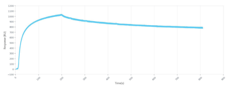
Understanding the signaling pathways that the biomarkers can activate can help in the development of checkpoint immunotherapies. Surface Plasmon Resonance (SPR) is a common technique used to identify and elucidate biomarker interactions, signaling pathways, mechanism of interaction, drug discovery and development of therapeutics by providing a label-free, real-time method of determining binding kinetics and binding affinity of key biomolecular interactions. Systems like Alto from Nicoya Lifesciences, the world’s first digital SPR instrument to integrate digital microfluidics and nanotechnology, can measure biomolecules in pM concentrations in various sample matrices with high specificity, while offering the versatility of working with a range of biomolecule types.
The following studies demonstrate the application of SPR bioassays in understanding and improving immunotherapies for breast and ovarian cancer.
Understanding the mechanism of immune escape
Gao et al., at Xijing Hospital in China’s Fourth Military Medical University, investigated the mechanism by which tumours in ovarian cancer can evade an immune attack by T cells, regulated by GDF-15. Growth differentiation factor-15 (GDF-15), a distal member of the secreted TGF-β superfamily, is expressed in dendritic cells (DCs) and is known to inhibit immune cell function thus limiting the effectiveness of immunotherapy.3 They identified a biomarker CD44, a single-chain non-kinase transmembrane proteoglycan, as a potential molecule on DCs that was found to be elevated in ovarian cancer patients. Immunoprecipitation combined with mass spectrometry, surface plasmon resonance, and co-immunoprecipitation assay were used to evaluate the interaction between GDF-15 and the surface molecules of DCs.
Using SPR, they found that GDF-15 bound to CD44 with high affinity with a KD of 1.98e-8 (±2.44e-11) M. Their results indicated that GDF-15 interacted with CD44, which suppressed the inhibitory effect of GDF-15, and in turn, facilitated immune escape. Thus, their study revealed a therapeutic target for immunotherapy against ovarian cancer.
Improving inefficiencies of monoclonal antibodies
Inhibition of human epidermal growth factor receptor 2 mediated cell signaling pathway is an important therapeutic strategy for HER2-positive cancers. To improve progression-free survival in breast cancer patients, combinatorial treatments of monoclonal antibodies and inhibitor drugs are commonly used. However, it is widely known that drugs selectively targeting tyrosine kinases (TKs) to treat breast cancer can cause resistance to inhibition, while monoclonal antibody therapeutics have high cost of production, high molecular weight, and are susceptible to enzymatic degradation.
Work done by Zheng et al., at The Chinese Academy of Medical Sciences demonstrated that a new class of peptidomimetics, γ-AApeptides, are low molecular weight, resistant to degradation and upon dimerization, form cyclic γ-AApeptides (M-3-6-D).4 They resemble the Y-shape antibody forming pseudo-loop formed by the heavy and light chains. With SPR, they showed that this cyclic antibody-like dimer enhanced the binding affinity of the ligand to the HER2 extracellular domain (ECD) with high affinity. Both M-3-6 and M-3-6-D showed a strong binding affinity to HER2 with a KD of 228 nmol/L and KD of 30 nmol/L respectively. This study successfully demonstrated that the γ-AApeptide-based artificial antibody specifically bound to HER2 and antagonized HER2 mediated cell signaling both in vitro and in vivo. Thus, the authors concluded that M-3-6-D was a promising antibody surrogate in anti-cancer therapeutics.
Advance your immunotherapy research with SPR
At Nicoya our benchtop SPR instruments have been innovated to improve the quality of research by providing label free, real-time binding kinetics and affinity information for a wide range of interactions, while being easy to use and accessible to every scientist.
To learn more about using SPR in your immunotherapy research, our application scientists would be happy to discuss your work in more detail.
References
- https://www.hopkinsmedicine.org/inhealth/about-us/immunotherapy-precision-medicine-action-policy-brief.html
- https://www.cancerresearch.org/en-us/news/2020/immunotherapy-drug-development-pipeline-growth
- Gao Y, Xu Y, Zhao S, et al. Growth differentiation factor-15 promotes immune escape of ovarian cancer via targeting CD44 in dendritic cells. Exp Cell Res. 2021;402(1):112522. doi: 10.1016/j.yexcr.2021.112522
- Zheng M, Chunpu L, Zhou M, et al. Peptidomimetic-based antibody surrogate for HER2. Acta Pharmaceutica Sinica B. 2021;11(9):2645-2654. doi: 10.1016/j.apsb.2021.04.016