[et_pb_section bb_built=”1″ admin_label=”section”][et_pb_row admin_label=”row” background_position=”top_left” background_repeat=”repeat” background_size=”initial”][et_pb_column type=”4_4″][et_pb_text admin_label=”Paragraph 1″ background_layout=”light” text_orientation=”justified” text_font_size=”16″ text_line_height=”1.9em” use_border_color=”off” custom_margin=”-25px|||” custom_padding=”||10px|” background_position=”top_left” background_repeat=”repeat” background_size=”initial” _builder_version=”3.0.96″]
Do you use NMR (nuclear magnetic resonance) to determine protein structure in your research? Dr. Michael Piazza, biochemistry PhD graduate from the University of Waterloo, uses both NMR and SPR (surface plasmon resonance) to obtain the key data needed for his recent publication. The research paper “Structural Consequences of Calmodulin EF Hand Mutations” published in Biochemistry studies calcium-dependent/independent properties of binding and activation of target proteins. Read our interview with Dr. Piazza to find out why NMR and SPR are the perfect pair when it comes to accelerating your research and publishing faster.
[/et_pb_text][/et_pb_column][/et_pb_row][et_pb_row background_position=”top_left” background_repeat=”repeat” background_size=”initial”][et_pb_column type=”4_4″][et_pb_text admin_label=”Paragraph 2″ background_layout=”light” text_font_size=”16″ text_line_height=”1.9em” use_border_color=”off” custom_margin=”-20px|||” background_position=”top_left” background_repeat=”repeat” background_size=”initial” _builder_version=”3.0.96″]
Tell us a bit about yourself and your research experience.
[/et_pb_text][/et_pb_column][/et_pb_row][et_pb_row][et_pb_column type=”2_3″][et_pb_text admin_label=”Paragraph 1″ background_layout=”light” text_orientation=”justified” text_font_size=”16″ text_line_height=”1.9em” use_border_color=”off” custom_margin=”-25px|||” custom_padding=”||10px|” background_position=”top_left” background_repeat=”repeat” background_size=”initial” _builder_version=”3.0.96″]
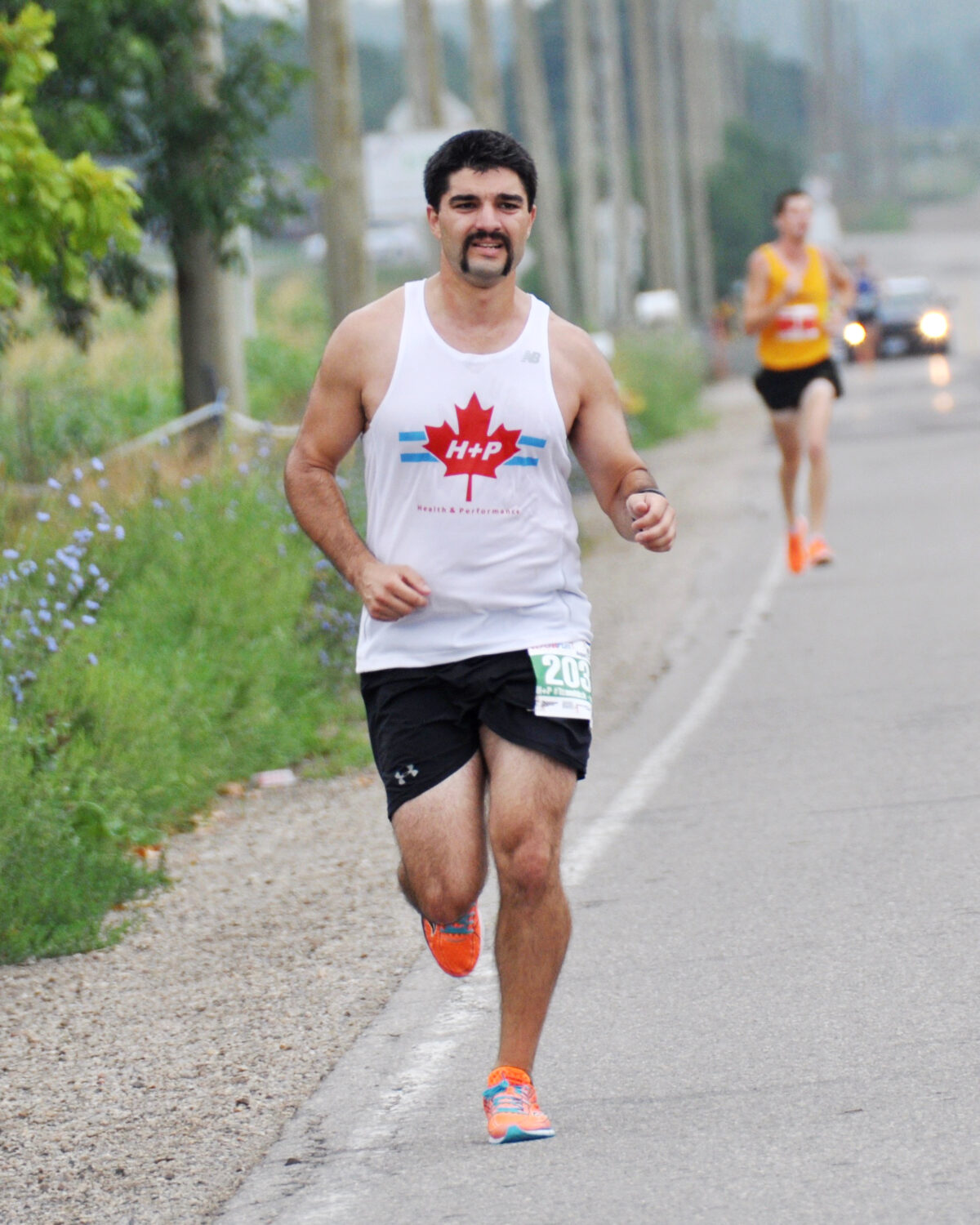
I recently completed my PhD in biochemistry at the University of Waterloo. My research focused on enzymes that are involved in producing nitric oxide, known as nitric oxygen synthase (NOS) enzymes, and their activation by calmodulin (CaM), a calcium-binding protein. Outside of my research, I also spend a lot of time running, playing and watching hockey and on the golf course. Sometimes I wonder if it’s tougher running a marathon or being a Maple Leafs fan.
[/et_pb_text][/et_pb_column][et_pb_column type=”1_3″]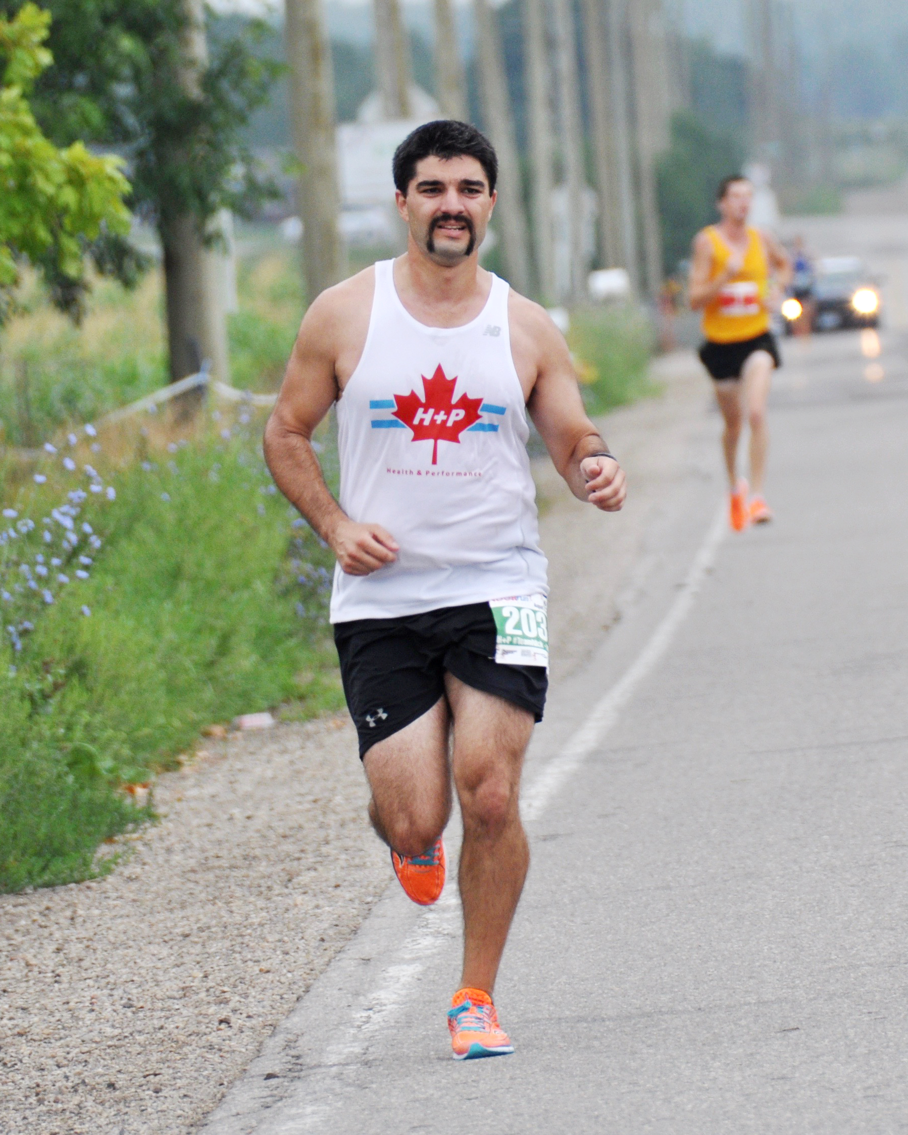 [/et_pb_column][/et_pb_row][et_pb_row admin_label=”row” background_position=”top_left” background_repeat=”repeat” background_size=”initial”][et_pb_column type=”4_4″][et_pb_text admin_label=”Paragraph 2″ background_layout=”light” text_font_size=”16″ text_line_height=”3em” use_border_color=”off” custom_margin=”-20px|||” background_position=”top_left” background_repeat=”repeat” background_size=”initial” _builder_version=”3.0.96″ header_line_height=”3em”]
Tell us about your recent publication “Structural Consequences of Calmodulin EF Hand Mutations” that uses OpenSPR.
[/et_pb_text][/et_pb_column][/et_pb_row][et_pb_row admin_label=”row” background_position=”top_left” background_repeat=”repeat” background_size=”initial”][et_pb_column type=”4_4″][et_pb_text admin_label=”Paragraph 1″ background_layout=”light” text_orientation=”justified” text_font_size=”16″ text_line_height=”1.9em” use_border_color=”off” custom_margin=”-25px|||” custom_padding=”||10px|” background_position=”top_left” background_repeat=”repeat” background_size=”initial” _builder_version=”3.0.96″]
All throughout literature, research groups have been using CaM mutant constructs that have their calcium-binding abilities knocked out to study the calcium-dependent/independent properties of binding and activation of target proteins by CaM. We investigated potential structural changes that these mutations may cause through the NMR structure determination of a fully calcium-deficient mutant construct and a complex with a target peptide that is known to interact with apoCaM. We then correlated the structural changes we observed with the binding kinetics determined using OpenSPR.
[/et_pb_text][/et_pb_column][/et_pb_row][et_pb_row background_position=”top_left” background_repeat=”repeat” background_size=”initial”][et_pb_column type=”4_4″][et_pb_text admin_label=”Paragraph 2″ background_layout=”light” text_font_size=”16″ text_line_height=”1.9em” use_border_color=”off” custom_margin=”-20px|||” background_position=”top_left” background_repeat=”repeat” background_size=”initial” _builder_version=”3.0.96″]
How did you use NMR to strengthen your publication?
[/et_pb_text][/et_pb_column][/et_pb_row][et_pb_row admin_label=”row” background_position=”top_left” background_repeat=”repeat” background_size=”initial”][et_pb_column type=”4_4″][et_pb_text admin_label=”Paragraph 1″ background_layout=”light” text_orientation=”justified” text_font_size=”16″ text_line_height=”1.9em” use_border_color=”off” custom_margin=”-25px|||” custom_padding=”||10px|” background_position=”top_left” background_repeat=”repeat” background_size=”initial” _builder_version=”3.0.96″]
NMR spectroscopy was used to determine the structure of a calcium-deficient CaM mutant and a CaM-mutant bound to the iNOS peptide. This allowed us to determine the structural changes these mutations caused and to show these calcium deficient CaM mutants may not be a true representation of apoCaM and may not allow for native-like interactions of apoCaM with its target protein.
[/et_pb_text][/et_pb_column][/et_pb_row][et_pb_row background_position=”top_left” background_repeat=”repeat” background_size=”initial”][et_pb_column type=”4_4″][et_pb_text admin_label=”Paragraph 2″ background_layout=”light” text_font_size=”16″ text_line_height=”1.9em” use_border_color=”off” custom_margin=”-20px|||” background_position=”top_left” background_repeat=”repeat” background_size=”initial” _builder_version=”3.0.96″]
Why was SPR important for your publication?
[/et_pb_text][/et_pb_column][/et_pb_row][et_pb_row admin_label=”row” background_position=”top_left” background_repeat=”repeat” background_size=”initial”][et_pb_column type=”4_4″][et_pb_text admin_label=”Paragraph 1″ background_layout=”light” text_orientation=”justified” text_font_size=”16″ text_line_height=”1.9em” use_border_color=”off” custom_margin=”-25px|||” custom_padding=”||10px|” background_position=”top_left” background_repeat=”repeat” background_size=”initial” _builder_version=”3.0.96″]
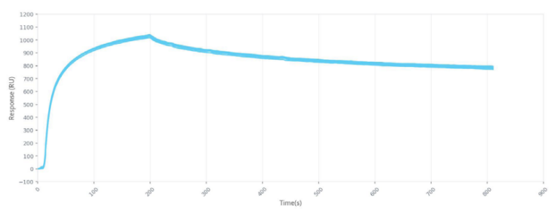
We used SPR to investigate whether the small structural differences between apoCaM and calcium-deficient CaM mutants result in appreciable changes in binding kinetics with the iNOS peptide at basal calcium concentrations. The differences in binding kinetics with CaM at basal calcium levels proved to be significant and provide further reason to scrutinize experiments that use calcium-deficient CaM mutants to represent the calcium-free form of the protein.
[/et_pb_text][/et_pb_column][/et_pb_row][et_pb_row background_position=”top_left” background_repeat=”repeat” background_size=”initial”][et_pb_column type=”4_4″][et_pb_text admin_label=”Paragraph 2″ background_layout=”light” text_font_size=”16″ text_line_height=”1.9em” use_border_color=”off” custom_margin=”-20px|||” background_position=”top_left” background_repeat=”repeat” background_size=”initial” _builder_version=”3.0.96″]
How did using both NMR & SPR help accelerate your research?
[/et_pb_text][/et_pb_column][/et_pb_row][et_pb_row admin_label=”row” background_position=”top_left” background_repeat=”repeat” background_size=”initial”][et_pb_column type=”4_4″][et_pb_text admin_label=”Paragraph 1″ background_layout=”light” text_orientation=”justified” text_font_size=”16″ text_line_height=”1.9em” use_border_color=”off” custom_margin=”-25px|||” custom_padding=”||10px|” background_position=”top_left” background_repeat=”repeat” background_size=”initial” _builder_version=”3.0.96″]
NMR spectroscopy allowed us to determine high resolution structures of these various CaM constructs and complexes to detect residue specific differences the mutations caused. While SPR was the key allowed us to correlate these structural differences through the acquisition of binding kinetic data. It allowed us to show that these mutations have consequences by providing more detail than just the binding affinity of CaM’s interaction with its target proteins through the OpenSPR’s ability to dive deeper into the interaction and provide association and dissociation rates.
[/et_pb_text][/et_pb_column][/et_pb_row][et_pb_row background_position=”top_left” background_repeat=”repeat” background_size=”initial”][et_pb_column type=”4_4″][et_pb_text admin_label=”Paragraph 2″ background_layout=”light” text_font_size=”16″ text_line_height=”1.9em” use_border_color=”off” custom_margin=”-20px|||” background_position=”top_left” background_repeat=”repeat” background_size=”initial” _builder_version=”3.0.96″]
Would you recommend other researchers using NMR to also use SPR? Why?
[/et_pb_text][/et_pb_column][/et_pb_row][et_pb_row admin_label=”row” background_position=”top_left” background_repeat=”repeat” background_size=”initial”][et_pb_column type=”4_4″][et_pb_text admin_label=”Paragraph 1″ background_layout=”light” text_orientation=”justified” text_font_size=”16″ text_line_height=”1.9em” use_border_color=”off” custom_margin=”-25px|||” custom_padding=”||10px|” background_position=”top_left” background_repeat=”repeat” background_size=”initial” _builder_version=”3.0.96″]
SPR is a great complement for NMR spectroscopy and I’d highly recommend it to any researchers that are using NMR to investigate interactions of biomolecule complexes. NMR is essential to probing any structural or dynamic effects of the interaction, however, SPR can take their investigation to another level. SPR would allow them to correlate their structural studies to specific binding kinetics, including more detailed information such as association and dissociation rates. It’s a pretty quick method for doing an experiment, and since it’s a label free interaction technique, there is no need to spend time and money labeling samples. The experiments can be optimized quite easily, and the binding conditions can be changed quick and easily to determine binding kinetics for your binding partners under a variety of conditions.
[/et_pb_text][/et_pb_column][/et_pb_row][et_pb_row admin_label=”row” make_fullwidth=”off” use_custom_width=”off” width_unit=”on” use_custom_gutter=”off” padding_mobile=”off” allow_player_pause=”off” parallax=”off” parallax_method=”off” make_equal=”off” parallax_1=”off” parallax_method_1=”off” column_padding_mobile=”on” background_position=”top_left” background_repeat=”repeat” background_size=”initial”][et_pb_column type=”4_4″][et_pb_cta button_url=”https://hubs.ly/H0fmXkC0″ url_new_window=”on” button_text=”Discuss your interactions with an application scientist today” use_background_color=”off” background_layout=”light” header_text_color=”#1e73be” body_font_size=”16″ use_border_color=”off” custom_button=”off” button_icon_placement=”right” background_position=”top_left” background_repeat=”repeat” background_size=”initial” _builder_version=”3.0.96″]